
Oncology centre
Our workplace is one of the modernly equipped workplaces providing radiation treatment. We provide treatment with linear accelerators, brachytherapy and therapeutic X-ray equipment. The preparation of radiation treatment is a demanding process and consists of several sequential steps. A team of physicians, physicists and radiology assistants are involved in the preparation of the radiation plan and the implementation of the radiation treatment.
The Radiology Centre has started operating a radiology department in July 2002. The computed tomography workplace was the first to be put into operation, followed by the magnetic resonance and ultrasound workplace. In 2011, the department underwent a significant upgrade with the replacement of the computed tomography scanner and the construction of a separate additional magnetic resonance department, for which a magnetic resonance machine with a magnetic field strength of 1.5 Tesla was purchased. At the turn of 2015 and 2016, the original and no longer suitable magnetic resonance machine was dismantled and replaced by a modern, technically very advanced machine with a magnetic field strength of 3 Tesla. At the same time as this innovation, a scanning workplace with indirect digitalisation (diagnostic X-ray) was built.
Contact us
The Oncology centre started operating the radiotherapy department in January 2003 and in March of the same year inaugurated a newly installed linear accelerator equipped with the latest technology for IMRT treatment. It continued to operate the Teragam cobalt irradiator, which was dismantled and replaced by a new linear accelerator in 2008.
Since 2006, the department has been equipped with a high dose rate brachytherapy (treatment from a short or contact distance) machine. The Wolf - Medizintechnik T-200 X-ray irradiator is used for the treatment of non-cancerous diseases. The department holds a quality certificate from the German company TÜV - Rheinland (quality certificate according to CSN EN ISO 9001:2016).
Spectrum of radiology examinations
Computed tomography - CT
The examination is carried out with the help of a Siemens Somatom Definition AS, which simultaneously scans 40 data layers. The instrument is specially designed for the purpose of radiation simulation, but this does not prevent its use for routine diagnostic examinations. The device can be used to examine any region of the body. This type of examination is most commonly used for examination of the chest, abdomen and pelvis.
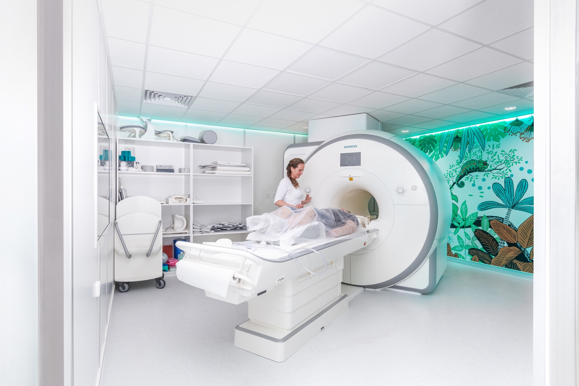
Magnetic resonance imaging
The examination is performed on a Siemens Magnetom Avanto (with a magnetic field strength of 1.5 T) and a Siemens Magnetom Skyra (with a magnetic field strength of 3 T). We offer a relatively wide range of MR examinations at our department, including MR examinations of the brain, spine, joints, abdominal and pelvic organs or MR angiography.
Ultrasound
The examination is performed on a Logiq 8 XDclear machine from GE Healthcare. The fully digital ultrasound system features unique technology for transmitting, receiving and processing the ultrasound signal, providing maximum image quality in all imaging modes.The system excels in ease of use, especially with its auxiliary colour LCD touch screen. The ultrasound features a 23" high resolution monitor and 4 electronically switchable active ports for connecting electronic probes. Probes are available for imaging abdominal organs, soft tissue, blood vessels, heart and examination of young children.
X-ray
Examinations are carried out on a Fomei Covator machine with indirect digital scanning.We are able to perform routine scans.
Oncology centre


Our satisfied patients
If you are still hesitant about the treatment in our medical facilities, listen to the stories of our satisfied clients.
Contact Us
Or simply leave us your phone number
and we will contact you back
 Download
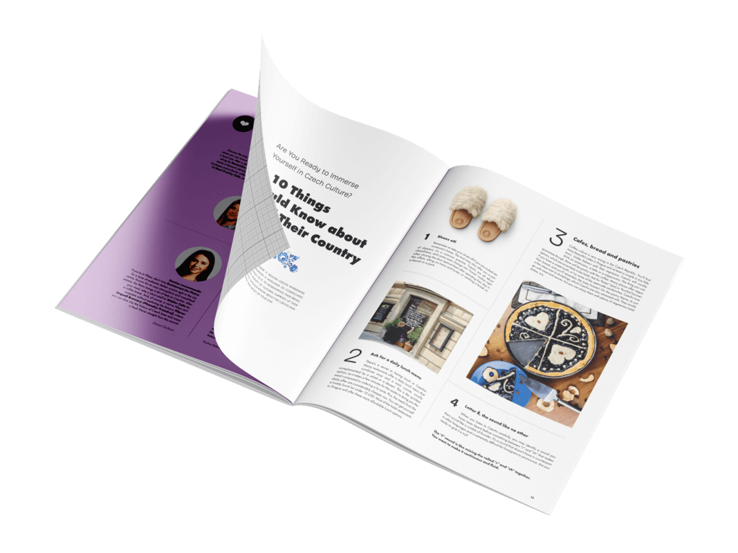
Discover the new Altoa Magazine!
Healthcare insights, our services, and fascinating stories about the Czech Republic.
Start reading